
Which Test Is Right for You: CT, MRI and X-Ray
- 6 minute read.
- Last Updated: 7/27/2026
When your doctor recommends imaging, it can be difficult to understand the differences between an X-ray, CT scan or MRI. While all three allow physicians to see inside the body, each has a unique purpose and provides different types of information.
Understanding these differences can help patients feel more informed and confident as they move through the diagnosis and treatment process.
Medical imaging plays a critical role in modern healthcare, helping providers identify injuries, detect disease, guide treatment plans and monitor progress over time. Across Covenant Health, advanced imaging services are available close to home. We offer patients access to the technology and expertise needed for accurate diagnoses and personalized care.

Why Imaging Matters Now
Medical imaging supports nearly every area of healthcare, including orthopedics, cardiology, oncology, women’s health, neurology and emergency medicine.
Often, imaging is one of the first steps in determining the cause of symptoms. It helps providers see what cannot be detected through a physical exam alone. This allows them to make informed decisions about treatment and next steps.
“One of the questions we hear most often is, ‘Why do I need this test instead of another one?'” says Margaret Wyatt, BSHCM, RT(R), CNMT, imaging services manager and radiation safety officer at Covenant Health’s Cumberland Medical Center. “Each imaging exam is designed to answer specific clinical questions. Choosing the right test helps physicians make an accurate diagnosis and create the most effective treatment plan.”
The three most common imaging exams are X-rays, CT scans and MRIs.
What Are X-Rays?
X-rays are one of the oldest and most widely used forms of medical imaging. They use a small amount of radiation to create images of structures inside the body, particularly bones and dense tissues.
Because they are quick and readily available, X-rays are often the first imaging test ordered to evaluate pain, injury or illness.
Best for:
- Broken bones
- Joint injuries
- Lung conditions, including pneumonia
- Certain infections
- Initial evaluations of pain or trauma
Benefits:
- Fast and efficient
- Widely available
- Typically completed in minutes
- Often used as a first step before more advanced imaging
“An X-ray gives us a quick look at what’s happening,” Wyatt explains. “If someone comes in after a fall or sports injury, we can often determine very quickly whether a fracture is present and whether additional imaging may be needed.”
What Are CT scans?
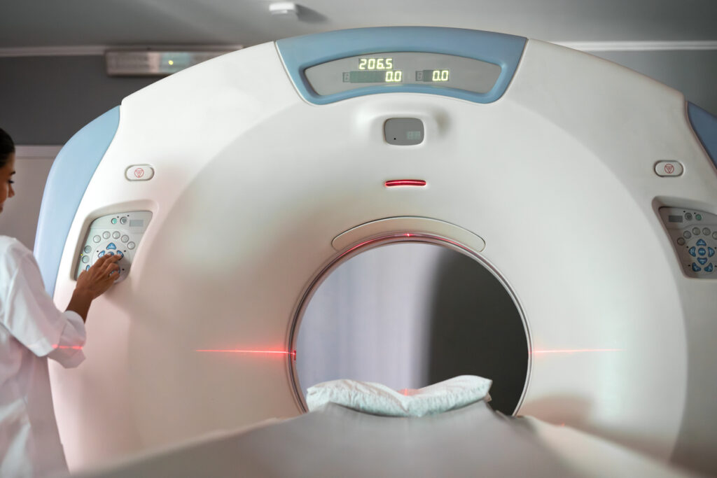
Computed Tomography (CT) scans use a series of X-ray images combined with sophisticated computer technology to create detailed cross-sectional views of the body.
Unlike traditional X-rays, CT scans show bones, organs, blood vessels and soft tissues in much greater detail. Physicians often rely on CT scans when they need a more complete picture of an injury, illness or internal condition.
Best for:
- Internal injuries
- Complex fractures
- Organ evaluation
- Stroke assessment
- Emergency conditions
- Certain cancer screenings and evaluations
Benefits:
- Highly detailed images
- Fast results
- Help guide treatment decisions
- Support surgical planning
“CT imaging is especially valuable when time matters,” says Wyatt. “In emergency situations, physicians can quickly evaluate internal injuries, bleeding or other serious conditions that may not be visible on an X-ray.”
What Are MRIs?
Magnetic Resonance Imaging (MRI) uses powerful magnets and radio waves to create highly detailed images without exposing patients to radiation.
MRI excels at visualizing soft tissues, making it one of the most effective tools for evaluating muscles, ligaments, tendons, nerves, the brain and spinal cord.
Best For:
- Brain and spine imaging
- Sports injuries
- Joint conditions
- Soft tissue evaluation
- Neurological disorders
- Certain cancer and organ assessments
Benefits:
- No radiation exposure
- Exceptional soft tissue detail
- Help detect conditions that may not appear on X-rays or CT scans
- Support precise diagnosis and treatment planning
“MRI allows us to see details that other imaging exams simply can’t provide,” Wyatt says. “When physicians need to evaluate a torn ligament, nerve compression or changes within the brain or spinal cord, MRI often gives the clearest answers.”
Because MRI images are extremely detailed, the exam typically takes longer than an X-ray or CT scan, but the additional information can be invaluable in diagnosing complex conditions.
Wyatt offers this simple comparison:
“Think of an X-ray as a snapshot, a CT scan as a flipbook and an MRI as a detailed painting. Each provides a different level of detail and answers different clinical questions. Choosing the right exam helps physicians get the information they need while avoiding unnecessary testing
Other Imaging Services Available
In addition to X-rays, CT scans and MRI, Covenant Health offers a wide range of imaging services designed to help diagnose, treat and monitor conditions throughout every stage of life.
Fluoroscopy
Fluoroscopy is a specialized type of X-ray that creates real-time moving images of structures inside the body. It is commonly used to evaluate swallowing and digestive disorders, guide certain procedures and assess movement within joints and organs.
Ultrasound
Ultrasound uses sound waves rather than radiation to create images. It is frequently used during pregnancy but also plays an important role in evaluating blood flow, abdominal organs, thyroid conditions and soft tissue abnormalities.
Mammography
Mammography is an essential breast cancer screening tool that can help detect abnormalities before symptoms develop. These tests improve opportunities for early detection and treatment.
Bone Density Testing (DEXA)
Bone density scans help assess bone strength and identify osteoporosis. This enables patients and providers to take steps to reduce fracture risk.
Nuclear Medicine
Nuclear medicine imaging uses small amounts of radioactive material to evaluate organ function and identify certain diseases. These commonly include heart conditions and some types of cancer.
“Every imaging exam has a specific purpose,” Wyatt shares. “The goal is always to match the right test to the patient’s symptoms, so physicians have the information needed to make informed decisions and provide the best possible care.”
When Should You Talk to Your Doctor About Imaging?
Your provider may recommend imaging if you experience:
- Sudden or severe pain
- A fall or sports injury
- Chest pain
- Trouble breathing
- Persistent headaches
- Swelling
- Joint pain
- Unexplained abdominal discomfort
Some groups may require imaging more frequently, including:
- Active children and teens
- Athletes
- Adults with chronic pain
- Older adults at risk for falls
- Patients monitoring known medical conditions
Convenient Imaging Close to Home
Covenant Health Imaging and Diagnostic Centers combine advanced radiological technology with compassionate, patient-centered care. With 11 locations across East Tennessee, patients have convenient access to a full range of imaging services, including X-ray, CT, MRI, ultrasound, mammography, bone density testing and more.
Learn more about Covenant Health Imaging and Diagnostic Centers and find a location near you.