Methodist Medical Center Neurodiagnostics
Understanding the Central Nervous System
Understanding the Central Nervous System
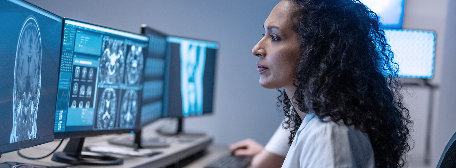
Modern imaging technology, especially magnetic resonance imaging (MRI), produces remarkably detailed views of the central nervous system. But images of the brain, spinal cord, nerve and muscles are not always enough when medical problems occur. Physicians need to know how the affected area is functioning. Neurodiagnostic tests provide that information.

The neurodiagnostics center at Methodist has a wide range of capabilities:
EEG: Electroencephalograms
Physicians use electroencephalograms to diagnose epilepsy, brain tumors, head injury, stroke, diseases that affect brain function, and other conditions. Several small metal disks are placed on the patient’s scalp and the patient is asked to follow simple directions, such as opening and closing their eyes or breathing faster than normal. This procedure, which is painless, records electrical activity in the brain and takes about an hour.
SSER: Somatosensory Evoked Response
Somatosensory evoked response tests may be ordered when a patient experiences back pain, difficulty walking, or pain, tingling or weakness in the arms or legs.Small metal disks are attached to the patient’s scalp and to various places along the path of the nerves being tested. The nerve is stimulated and the computer records the brain’s response to the stimulus. SSER tests take about an hour.
VER: Visual Evoked Response
Visual evoked response testing helps physicians diagnose problems with nerves that affect sight. It may be useful when patients have double vision, decreased or loss of vision, or headaches. Small metal disks are placed on the scalp and the patient is asked to watch a pattern flash on a screen for about a minute. A computer records electrical responses from the patient’s brain.
NCS: Nerve Conduction Studies
Nerve conduction studies give physicians important information about nerve and muscle function. They help identify, for example, whether a patient’s problem is in the spinal cord, muscles, nerves leading to muscles, or other structures in the body. Small discs or electrodes are placed on the patient’s limbs, the nerves are stimulated, and the responses are recorded.
IOM: Intraoperative Neurophysiological Monitoring
Intraoperative neurophysiological monitoring assesses a patient’s central nervous system during certain types of surgery, including brain surgery, orthopedic surgeries of the spine and procedures involving the heart, brain and blood vessels.
TCD: Transcranial Doppler
Transcranial doppler is a study of the blood flow of the brain that provides useful information to the doctor for those patients experiencing strokes (CVAs) or mini-strokes (TIAs).
About 50 million Americans suffer from conditions involving the central nervous system. These conditions include:
- Huntington’s disease
- Muscular dystrophy
- Other genetic diseases
- Degenerative conditions in adults such as Parkinson’s disease and Alzheimer’s
- Infectious diseases including dementia related to AIDS
- Developmental problems such as cerebral palsy
- Brain tumors
- Stroke
- Epilepsy
- Head and spinal cord injuries
